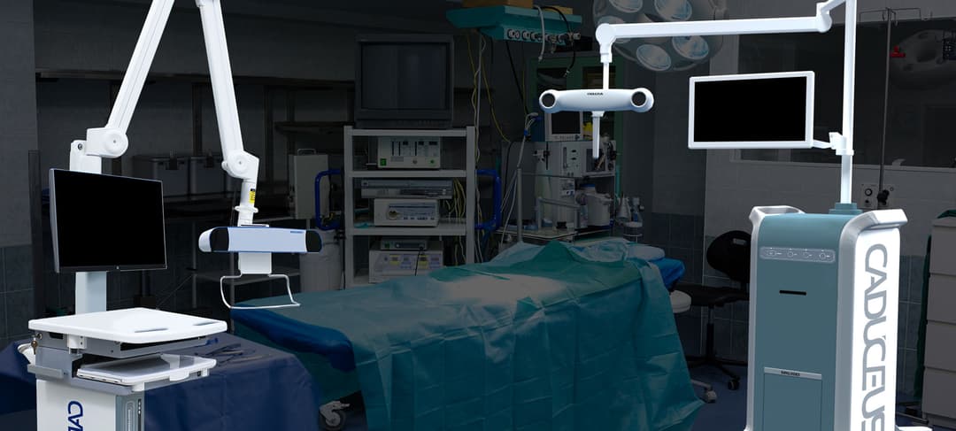
How it Works
During surgery, a CT scan is performed on the patient while on the operative table and transferred to a computer to create a detailed, three-dimensional virtual map of their spine. An optical tracking system follows the surgeon’s instruments and projects their exact position onto the 3D model on a monitor. This allows the surgeon to see the instrument's precise location in relation to the patient's anatomy, including vital structures like the vertebrae, nerves and blood vessels, with a high degree of accuracy.
Benefits
This technology significantly enhances surgical precision, which is particularly crucial for complex procedures like placing pedicle screws in spinal fusions or performing osteotomies in spinal deformity patients. By providing real-time guidance, computer navigation helps minimize the risk of complications, reduces the need for repeat x-ray radiation throughout the procedure, and can allow for minimally invasive surgeries. This often translates to a safer operation, shorter hospital stays, and a faster recovery for the patient.